
Since the invention of light microscopy in the 17th century, when the compound microscope has enabled a foretaste of cellular structures on a very small scale of micrometers, the evolution of techniques in light microscopy has gone hand-in-hand with new scientific discoveries in the life and material sciences. The diffraction limit of the light microscope has been circumvented by the fluorescence light microscopy technique. The super-resolution visible light microscopy though offers access to the nanoscale, but it also imposes the diffraction limitation by the wavelength of visible light. This is resolved by electron microscopy, which allowed smaller wavelengths in the sub-atomic limits. The biggest disadvantage of modern electron microscopes is their small penetration depth for samples measuring numerous micrometers in breadth.
Ptychography with brilliant X-rays was developed in the late 2000s as a technique that eventually combines Scanning Transmission X-ray microscopy (STXM) with Coherent Diffraction Imaging (CDI) has changed the game altogether.

First time in the history of science, we can now accurately measure the mass of human chromosomes using a powerful X-ray source at the UK’s national synchrotron science facility, the Diamond Light Source to determine the individual masses of all 46 chromosomes in human cells. The newly discovered masses are higher than previously expected, that is 20 times the mass of DNA, which hints towards the other unknown mass that could be hiding inside, and yet to be explored.
Ian Robinson, a biophysicist from the University College London said “Our measurement suggests the 46 chromosomes in each of our cells weigh 242 picograms (trillionths of a gram). This is heavier than we would expect, and, if replicated, points to unexplained excess mass in chromosomes.”
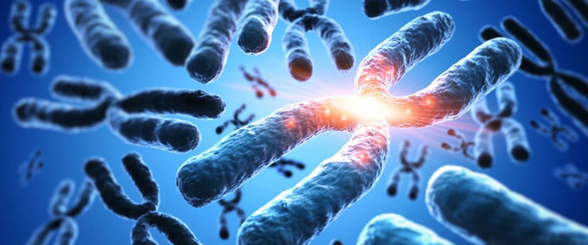
Chromosomes are thread-like structures that contain DNA, which in turn has genetic instruction. Humans have 23 pairs of chromosomes, out of which one is a sex chromosome. Though discovered in the 19th century, a lot about chromosomes is yet to be discovered. The new method of hard X-ray ptychography will allow scientists to better understand it.
X-ray ptychography uses a particle accelerator known as a synchrotron, to produce a strong beam of X-rays, which when passes through chromosomes, creates an interference pattern by diffraction that allows scientists to create a high-resolution 3D model of the same chromosome. For the study purpose, these researchers used the human White blood cells at metaphase and were able to calculi the number of electrons, and electron density of the chromosome.
The researchers have found an unexpected mass of the chromosomes and aren’t sure, what’s causing it to be this high, but the answer could be of creating an advantage to science, in better understanding our body’s mechanisms and diseases. Archana Bhartiya a Bioscientist believes that an improved understanding of chromosomes can have significant consequences for human health. For example, diagnostics for cancers involves rigorous study of the chromosomes, and any developments in our abilities to picture chromosomes would be of great value.


Add comment